 Southwest Florida's Health and Wellness Magazine Health and Wellness Articles

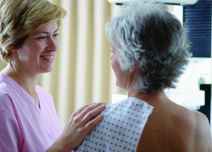 Now women have an imaging program designed specifically to meet their needs with the latest technology and professional, responsive care – at Naples Diagnostic Imaging Centers.
Breast Ultrasound
NDIC performs breast ultrasound as a part of a multi-modality approach to breast care. Ultrasound, also know as sonography, uses high-frequency sound waves (above the hearing range) to produce images of the breast tissue. The various densities of the tissues within the breast (including glandular, fibrous, connective and fatty elements) reflect different amounts of the sound waves to produce an image that has various shades of gray that correlate to the densities of the tissues. Breast ultrasound is used to evaluate breast masses that are felt and/or demonstrated by mammography or breast MRI to determine if the masses are cystic (fluid-filled) or solid. Breast ultrasound imaging can aid in differentiating benign from malignant processes, assessing breast implant problems and diagnosing and monitoring breast inflammation or abscess formation as well as providing guidance of need placement for aspiration or biopsy.
Mammography
Doctors recommend that all women over 40 have an annual mammogram as a preventative measure; an earlier start is recommended for those with a family history or at high risk for the disease. When women choose NDIC for their mammography, they can find even greater peace of mind. That’s because screening mammograms at NDIC use computer-aided detection (CAD) to alert physicians to potential abnormalities. Studies show nearly 25% of breast cancer would be detected earlier using CAD.
Direct contact with interpreting physicians is also available, and a patient may choose to discuss her test results with the radiologist immediately upon completion of her mammogram. No physician referral is required for their test.
Digital Mammography with Computer
Aided Detection
Now only at NDIC, digital mammography has replaced old film technology – providing quicker, clearer and more reliable information and faster more comfortable exams, too. Even more, the system uses Computer-Aided Detection, technology that highlights any abnormalities it detects, and alerts the radiology physician to focus on these areas. As a result, 23.4 % more cancers can be detected earlier, at a more treatable stage.
Stereotactic (Mammographically Guided)
Breast Biopsy
Mammography and other breast imaging cannot prove that an abnormal area of breast tissue is cancer. If mammography or another breast imaging modality raises significant suspicion of cancer, tissue must be removed for examination under a microscope to tell if it is cancer with certainty. This can be done by a breast biopsy.
The staff and Radiologist at NDIC are among the best at taking care of this sensitive subject and will assist you in any way to provide you with outstanding care and comfort.
Breast MRI with Computer Aided Detection
This breakthrough in women’s imaging can save patients from undergoing stereotactic biopsy or invasive surgical procedures, and also can track the effectiveness of treatment. A painless diagnostic technique, the MRI actually enables physicians to designate a highly-specific area for examination. As a result, the physician has an extremely precise image of medical conditions without invasive procedures or radioactive exposure.
Then, computer-aided detection harnesses the power of computers to allow for increased sensitivity in interpreting the results, alerting the radiology physician to focus on any suspicious areas. This technology is particularly useful for women with high risk for breast cancer or with dense breasts.
Breast-Specific-Gamma-Imaging (BSGI)
BSGI is a new and specialized breast imaging technique that incorporates the uses of Nuclear Medicine with breast imaging. BSGI is very useful in women with dense breast tissue, suspicious areas on a mammogram, lumps that can be felt but not seen on mammography or ultrasound, breast with implants and augmentation, and scarring from previous surgeries.
NDIC offers this new technique in detecting breast abnormalities. BSGI can aid in diagnosis when mammography is inconclusive revealing important information that can help your doctor more accurately determine if an area of concern is cancerous or not.
Physician Spotlight
Dr. Theresa Vensel is certified by the American Board of Radiology and specializes in diagnostic radiology and women’s imaging. After receiving her B.A. from the University of Virginia in 1996, she attended the University of Miami School of Medicine where she received her M.D. In 2000. Dr. Vensel’s sincere and compassionate care set her apart from many others in her field.
239-593-4200
www.naplesxray.com