 Southwest Florida's Health and Wellness Magazine Health and Wellness Articles

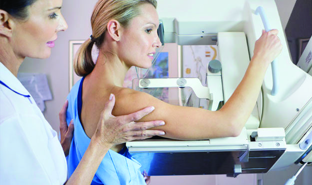 Mammography or a mammogram is an x-ray examination of the breast tissue. Mammography has been used for years to detect and diagnose breast diseases in both women and men. While most mammography is done on a screening basis, it is crucial in diagnosing the cause of breast symptoms such as breast lumps, breast pain, and nipple discharge.
Annual screening mammograms are performed on patients who are asymptomatic (has no breast complaints) and meet the criteria for a mammogram. These criteria are generally age (over the age of 40), and family history of breast cancer.
Screening Mammography
The goal of annual screening mammograms is to detect breast cancer when it is still too small to be felt by either the woman or her physician. Early detection of small breast cancers by screening mammography dramatically increases the chances for successful treatment of the disease. In the U.S., one out of eight women will develop breast cancer during her life. The earlier the disease is diagnosed, the higher the chances of a complete cure. When breast cancer is detected in the localized stage without having spread to the lymph nodes, the five year survival rate is 98%. If the cancer has spread regionally to the axillary (underarm) lymph nodes the rate drops to 76%.
If you experience unusual tenderness, pain, nipple discharge or notice a lump in your breast (even if you are in your early twenties), contact your personal physician immediately and come in for a diagnostic evaluation. The best treatment for breast disease is early detection.
• Women 20 years of age and older should perform breast self-examinations monthly
• Women 20-39 should have a physical examination of the breast every three years, performed by a health care professional such as a physician.
• Women 40 and older should have a physical examination of the breast every year.
• Women 40 and older should have a mammogram every year.
Diagnostic Mammography
Diagnostic mammograms are performed on patients with a breast complaint (for example, a breast lump, pain or nipple discharge etc.) or have had an abnormal screening mammogram. During a diagnostic mammogram, additional imaging of a breast abnormality will be taken and carefully evaluated. These images may include coned view with magnification which increases the appearance of an abnormality and makes it easier to see. The additional views to be obtained are tailored to the patients needs by the Radiologist. During this visit the Radiologist will come and speak with you about your results.
A complete diagnostic work up may show that a lesion or area of abnormal tissue has a high likelihood of being benign (not cancer), and although the Radiologist may believe that the chances of this area developing into cancer are low, you may be asked to have the area re-imaged in a few months. Or the diagnostic mammogram may show that the area is not of concern and you will then be instructed to continue your routine screening mammograms.
Finally a diagnostic mammogram may suggest that a biopsy is needed to tell whether or not a lesion is cancer. Mammography cannot prove that an abnormal area is cancer. If mammography raises a significant suspicion of cancer, tissue must be removed for examination under a microscope to tell if it is cancer with certainty. This can be done by needle biopsy or open surgical biopsy. A recommendation of biopsy does not necessarily mean that the abnormality is cancer. About 70% of all breast lesions that are evaluated with biopsy are found to be benign.
How Do I Prepare for a Mammogram?
When you arrive for you to have your mammogram, it is preferable that you dress in a two-piece outfit, since you will be asked to remove all clothing from the waist up. Do not use any lotions, powders, perfumes, or deodorant in the breast or underarm area the day of your test (particles from the chemicals can distort the images). If you are still of reproductive years, try to avoid scheduling your exam the week before your period, as your breasts will be tender. If possible, obtain prior mammograms and make them available to the imaging facility before your appointment. The use of comparison to prior images is very valuable.
You can feel comfortable in having your mammogram performed at Naples Diagnostic Imaging Center. The Radiologists are highly trained and are respected in the field of mammography. We perform thousands of mammograms annually and pride ourselves in helping women detect breast disease. We use only state-of-the-art equipment and are certified and accredited by the American College of Radiology and the FDA.
To learn more about Mammography, and other imaging services available at NDIC call 239-593-4222.