 Southwest Florida's Health and Wellness Magazine Health and Wellness Articles

 A variety of imaging techniques may be performed to determine the size, shape, location, and consistency of the ovaries.
Imaging tests like computed tomography (CT) scans, magnetic resonance imaging (MRI) scans, and ultrasound studies can confirm whether a pelvic mass is present. These diagnostic studies may also be useful if your doctor is looking to see if ovarian cancer has metastasized to other tissues and organs.
Ultrasound
Ultrasound (ultrasonography) is the use of sound waves to create an image on a video screen. Sound waves are released from a small probe placed in the woman’s vagina or on the surface of her abdomen. The sound waves create echoes as they enter the ovaries and other organs. The same probe detects the echoes that bounce back, and a computer translates the pattern of echoes into a picture.
Ultrasound is often the first test done if a problem with the ovaries is suspected. It can be useful in finding an ovarian tumor and seeing if it is a solid mass or a fluid-filled cyst. It can also be used to get a better look at the size of the ovaries and the internal appearance or complexity. These factors help the doctor decide which masses or cysts are more worrisome.
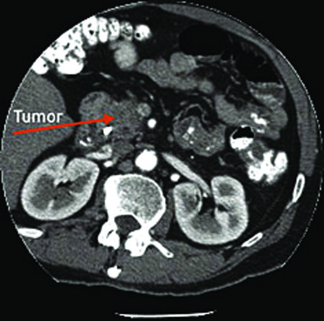
Computed tomography (CT) scans
The CT scan is an x-ray procedure that produces detailed cross-sectional images of your body. Instead of taking one picture, like a conventional x-ray, a CT scanner takes many pictures as it rotates around you. A computer then combines these pictures into an image of a slice of your body. The machine will take pictures of multiple slices of the part of your body that is being studied.
A CT scanner has been described as a large donut, with a narrow table in the middle opening. You will need to lie still on the table while the scan is being done. CT scans take longer than regular x-rays, and you might feel a bit confined by the ring while the pictures are being taken.
CT scans do not show small ovarian tumors well, but they can see larger tumors, and may be able to see if the tumor is growing into nearby structures. A CT scan may also find enlarged lymph nodes, signs that the cancer has spread to the liver or other organs, and if an ovarian tumor is affecting your kidneys or bladder.
You may be asked to drink 1 to 2 pints of a liquid before the CT scan called oral contrast. You might also receive an IV (intravenous) line through which a different kind of contrast dye is injected. Contrast dyes help better outline the structures in your body.
The injection can cause some flushing or a redness and warm feeling that may last hours to days. A few people are allergic to the dye and get hives. Rarely, more serious reactions like difficulty breathing and low blood pressure can occur. Medicine can be given to prevent and treat allergic reactions. Be sure to tell the doctor if you have had a reaction to any contrast material used for imaging testing or are allergic to Iodine.
CT scans are not usually used to biopsy an ovarian tumor, but they can be used to biopsy a suspected metastasis. For this procedure, called a CT-guided needle biopsy, the patient stays on the CT scanning table, while a radiologist moves a biopsy needle toward the location of the mass. CT scans are repeated until the doctors are confident that the needle is within the mass. A fine needle biopsy sample (tiny fragment of tissue) or a core needle biopsy sample (a thin cylinder of tissue about ½ inch long and less than 1/8 inch in diameter) is removed and examined under a microscope.
Magnetic resonance imaging (MRI) scans
MRI scans use radio waves and strong magnets to make pictures of organs within the body. The energy from the radio waves is absorbed and then released in a pattern formed by the type of tissue and by certain diseases. A computer translates the pattern of radio waves given off by the tissues into a very detailed image of parts of the body. Not only does this produce cross-sectional slices of the body like a CT scanner, it can also produce slices that are parallel with the length of the body. Contrast material might be injected into a vein (same as with a CT scan). Even though MRI scans are rarely used to look for ovarian cancer, sometimes they can be beneficial.
MRI scans usually take longer than CT scans. You will be placed inside a tube, which is confining and can be difficult for people with claustrophobia. The machine may make a thumping noise that can be disturbing.
Chest x-ray
A Chest x-ray may be done to determine whether ovarian cancer has spread to the lungs. Tumors that have spread to the lungs may cause fluid to collect around the lungs. This fluid, called a pleural effusion, can be seen with chest x-rays as well as other types of scans.
Positron emission tomography (PET) scan
In this test, which can be more definitive when combined with a CT scan (PET/CT scan) uses radioactive glucose to look for the cancer. Because cancers use glucose at a higher rate than normal tissues, the radioactivity will tend to concentrate in the cancer. A scanner can spot the radioactive deposits. PET scans can helpful in finding small collections of cancer when it has spread to other parts of the body.
Each of these and other types of diagnostic imaging services are available at NDIC. To schedule a screening or to learn more about a specific imaging service, call 239-593-4200 today.
NDIC
239-593-4222
www.NaplesImaging.com