 Southwest Florida's Health and Wellness Magazine Health and Wellness Articles

By Heidi Smith, Contributor
Women in the Venice area who have been diagnosed with breast cancer or who are at high risk for breast cancer now have access to the latest breast MRI (magnetic resonance imaging) technology to aid in further diagnosis and screening through Venice Regional Bayfront Health.
Venice Regional’s new breast MRI technology uses radio waves and strong magnets to make detailed pictures of the inside of the breast that are then viewed by the radiologist using an advanced imaging system. The technology may be used to help determine the extent of previously diagnosed breast cancer or to screen for breast cancer, explained Melinda B. Hart, M.D., a breast surgeon and an independent member of the medical staff at Venice Regional.
“Women at high risk for breast cancer or those who have had an abnormal mammogram should not delay in following up on the care plan recommended by their physician,” said Dr. Hart.
For women who already have been diagnosed with breast cancer, breast MRI may be used to help measure the size of the cancer, look for other tumors in the breast, and to check for tumors in the opposite breast. But not every woman who has been diagnosed with breast cancer needs a breast MRI. According to the American Cancer Society, MRI is not recommended as a screening test by itself because it can miss some cancers that a mammogram would find.
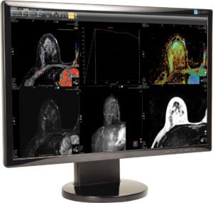
How It Works
At Venice Regional, the breast MRI is an outpatient procedure. As with all activities at the hospital’s facilities, strict protocols are in place to protect patients and staff from infectious diseases, such as COVID-19. These procedures are explained to patients when scheduling appointments.
Just as mammograms are done using x-ray machines specially designed for the breasts, breast MRI also requires special equipment called breast coils. At Venice Regional, the images collected by the breast MRI are sent to a special display unit that allows the radiologist to review the images with greater clarity and accuracy, and potentially, earlier detection of cancer.
Before the test, the technologist will ask if you have any types of metal in your body. If you have any of these types of medical implants, you should not even enter the MRI scanning area unless you’re told it’s okay to do so by a radiologist or technologist: an implanted defibrillator or pacemaker; clips used on a brain aneurysm; a cochlear (ear) implant; or metal coils inside blood vessels.
You’ll be asked to undress and put on a gown or other clothes without zippers or metal. Be sure to remove any metal objects you can, like hair clips, jewelry, dental work, and body piercings. The test is given with intravenous contrast so a technologist will start an IV before the procedure.
For the scan, you’ll lie face down on a narrow, flat table. Your breasts will be positioned through an opening in the table (the breast coil) so they can be scanned without being compressed. The technologist may use pillows to make you comfortable and help keep you from moving. The table then slides into a long, narrow tube. The test is painless, but you have to lie still inside the narrow tube. You may be asked to hold your breath or keep very still during certain parts of the test.
If you’re uncomfortable in confined spaces, your physician may prescribe medication to help you relax. The machine makes loud, thumping, clicking, and whirring noises, much like the sound of a washing machine, as the magnet switches on and off. The technologist can give you earplugs or headphones to help block out noise during testing.
You’ll be in the exam room alone, but you can talk to the technologist, who can see and hear what’s going on. As the scanning begins, the technologist will inject a contrast dye through an IV in your arm. The dye helps to clearly show breast tissue details. Let the technologist know if you have any kind of allergies or have had problems before with any contrast or dye used in imaging tests.
It’s important to stay very still while the images are being made. Each set of images usually takes a few minutes, and the whole test usually takes between 45 and 60 minutes. After the test, you may be asked to wait while the pictures are checked to see if more are needed.
“Breast MRI — in addition to mammograms — can be an important tool in managing the health of women with certain high-risk factors for breast cancer,” Dr. Hart said. “Talk with your doctor about your family and medical history to understand your risk factors and which screening procedures are best for you.”
To learn more about breast MRI, speak with your doctor or call 941-483-7205.
Venice Regional Bayfront Health
Call 941.483.7205 or visit VeniceRegional.com